Most of us do not think about eye pressure until a physician points it out at a routine checkup. What if a condition that could threaten your vision was developing right now, but you had no way or feeling of it developing? That’s exactly what makes Ocular hypertension concerning.
Your eyes maintain a delicate balance of pressure everyday. When that pressure becomes too high a condition called ocular hypertension is developed. It may not affect your vision immediately, but it can slowly become a serious concern over time if not treated early.
What is ocular hypertension?
Ocular hypertension is high intraocular pressure or high pressure inside your eyes. Normal eye pressure is between 11 to 21 mmHg. When the range of pressure is above the normal range with no detectable changes to the vision that is when ocular hypertension occurs.
It is prevalent among the people of all age groups, but it occurs frequently in African Americans, individuals over 40 and individuals with a hereditary condition of glaucoma and ocular hypertension. Bilateral ocular hypertension happens in both the eyes. Whereas, unilateral hypertension is the one that occurs only in one eye.
Ocular hypertension symptoms
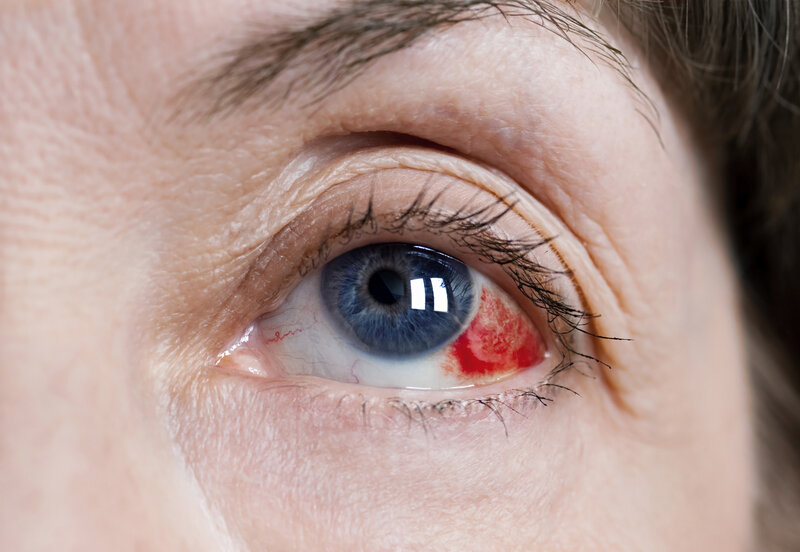
Ocular hypertension is a silent condition and there are typically no symptoms. This is the reason why regular eye examinations are necessary. In some individuals when there is elevated eye pressure, they can experience:
- Eye pain or discomfort
- Hazy or blurry vision
- Headache especially near eyes
- Seeing Halos around eyes
Few of the questions that you can ask your healthcare provider during eye test are:
- How high is my eye pressure?
- Has my optic nerve been damaged?
- Is my peripheral vision normal?
- Is treatment necessary?
- Are there any signs of eye damage due to an injury?
What causes Ocular Hypertension?
The most common cause of ocular hypertension is the subtle reduction in the eye’s ability to drain aqueous humour through its natural drainage ability called the trabecular meshwork. Let me give you a simple explanation:Imagine there is a water balloon. The more water you will fill in the balloon, the more pressure will be built in it. The same is the case with eye pressure. Causes of the build up are:
- The drainage angle is closed
- The anterior chamber is closed but not draining properly
- Pigment or protein flakes are blocking the drainage angle
- Eye cancer
- Previous eye damage
Recent studies have also indicated that stress can increase intraocular hypertension.
Risk factors of Ocular hypertension
There are several risk factors that affect ocular hypertension. Few of the risk factors cannot be changed like age, hereditary conditions, ethnicity or other medical conditions. The other risk factors are:
- Hypertension
- Hypotension
- Diabetes
- Extreme near-sightedness i.e. myopia
- Thin central cornea
People with thinner than average corneas have a higher risk of developing ocular hypertension.
- Bleeding at the optic nerve head
- Pigment dispersion syndrome
Pigment dispersion syndrome is a condition in which the pigment from the coloured part of the eyes flakes off and obstructs the draining system.
- Long-term steroid use
Extended use of steroid medications including eye drops, inhalers, creams and oral steroids can increase the risk.
Ocular hypertension vs Glaucoma
Ocular hypertension and glaucoma both involve elevated eye pressure but they differ significantly in the way they impact on eye health. Ocular hypertension refers to high intraocular pressure without any detectable damages to the optic nerve or vision loss. On the other hand, glaucoma is the elevated intraocular pressure along with damage to the optic nerve and can lead to permanent vision loss if untreated. Ocular hypertension is just a cause of glaucoma and not everyone who has it will develop the disease.
Who is most at risk?
Certain groups are more likely to develop ocular hypertension:
- Individuals above 40 years
- People with a family history of glaucoma
- Individual with diabetes
- Long-term steroid users
- People with extreme near-sightedness
- Individuals with thin corneas
- People with previous eye injuries
Diagnosis
Since symptoms are usually absent, diagnosis of the condition is totally dependent on the following eye examinations:
- Tonometry
Tonometry is the primary test to measure intraocular pressure. It is the first indication of elevated eye pressure In this test, the physician numbs the eyes and uses an instrument to measure the cornea that resists pressure.
- Pachymetry
Pachymetry is a quick, painless technique to measure the thickness of the cornea. As corneal thickness is the one that helps the physicians to detect the thickness of cornea and interpret intraocular pressure and also determine the risk of glaucoma.
- Gonioscopy
Gonioscopy is the test that checks whether the eye’s drainage system is working properly or not. In this test, a special contact lens is used.
- Optic nerve evaluation
Ocular hypertension is the major risk factor for glaucoma and thus physicians inspect the optic nerves. They may use advanced imaging for detecting the early signs of optic nerve damage.
- Visual field test
A test to check your peripheral vision to ensure the elevated pressure has not caused any vision loss.
Regular monitoring helps to keep a record of ocular hypertension if it remains stable or progresses towards glaucoma.
Can Ocular Hypertension cause blindness?
Ocular hypertension won’t itself cause blindness but elevated eye pressure can cause optic nerve damage which connects the eye to the brain. High eye pressure physically restricts or compresses blood flow to the optic nerve. This nerve damage destroys the delicate nerve fibers, which initially causes blind spots on the peripheral vision. If left untreated, the condition can aggravate and lead to blindness.
Early intervention dramatically reduces the risk of future complications.
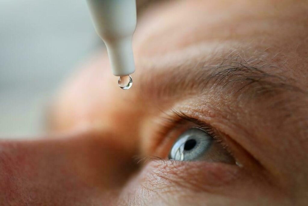
Ocular hypertension treatment
Eye drops like Dorzox T 5 ml are often the first line of treatment when it comes to treating ocular hypertension. They work by reducing the fluid production, improving fluid drainage and lowering intraocular pressure. Consistent use of the eye drops can significantly reduce the risk of glaucoma development. Laser treatments: For some conditions and patients, laser treatments may improve fluid drainage and lower eye pressure.
If the risk of glaucoma is low, your eye specialist may recommend regular follow-up appointments to monitor pressure and optic nerve health.
Final Thoughts
Ocular hypertension is a silent eye condition that has symptoms that are unnoticed. While it may not immediately affect vision but if left untreated it can affect the vision and cause complete blindness. It’s important for everyone to have regular eye exams, follow the medications provided by the healthcare provider.
When it comes to protecting your eyes, prevention and early action are far better than waiting for symptoms to appear. Timely intervention will protect your eyes.
FAQs
1. What is ocular hypertension?
Ocular hypertension is the condition that occurs when the pressure in the eye exceeds the normal pressure in the eye.
2. Do ocular hypertension always lead to glaucoma?
No. Ocular hypertension does not always lead to glaucoma. According to studies, only 9.5% of the patients progressed towards glaucoma.
3. Can ocular hypertension be cured?
Ocular hypertension does not have a permanent cure, but it can be prevented by using eye drops and laser therapy.
4. Is ocular hypertension dangerous?
Ocular hypertension may not cause immediate harm. However, if not treated eye pressure can increase the risk of glaucoma and vision loss.